Nondestructive Weld Examination
The ABC's of Nondestructive Weld Examination
Reprinted courtesy of Welding Journal magazine
An understanding of the benefits and drawbacks of each form of nondestructive examination can help you choose the best method for your application.
The philosophy that often guides the fabrication of welded assemblies and structures is "to assure weld quality." However, the term "weld quality" is relative. The application determines what is good or bad. Generally, any weld is of good quality if it meets appearance requirements and will continue indefinitely to do the job for which it is intended. The first step in assuring weld quality is to determine the degree required by the application. A standard should be established based on the service requirements.
Standards designed to impart weld quality may differ from job to job, but the use of appropriate weld techniques can provide assurance that the applicable standards are being met. Whatever the standard of quality, all welds should be inspected, even if the inspection involves nothing more than the welder looking after his own work after each weld pass. A good-looking weld surface appearance is many times considered indicative of high weld quality. However, surface appearance alone does not assure good workmanship or internal quality.
Nondestructive examination (NDE) methods of inspection make it possible to verify compliance to the standards on an ongoing basis by examining the surface and subsurface of the weld and surrounding base material. Five basic methods are commonly used to examine finished welds: visual, liquid penetrant, magnetic particle, ultra-sonic and radiographic (X-ray). The growing use of computerization with some methods provides added image enhancement, and allows real-time or near real-time viewing, comparative inspections and archival capabilities. A review of each method will help in deciding which process or combination of processes to use for a specific job and in performing the examination most effectively.
Visual Inspection (VT)
Visual inspection is often the most cost-effective method, but it must take place prior to, during and after welding. Many standards require its use before other methods, because there is no point in submitting an obviously bad weld to sophisticated inspection techniques. The ANSI/AWS D1.1, Structural Welding Code - Steel, states, "Welds subject to nondestructive examination shall have been found acceptable by visual inspection." Visual inspection requires little equipment. Aside from good eyesight and sufficient light, all it takes is a pocket rule, a weld size gauge, a magnifying glass, and possibly a straight edge and square for checking straightness, alignment and perpendicularity.
Before the first welding arc is struck, materials should be examined to see if they meet specifications for quality, type, size, cleanliness and freedom from defects. Grease, paint, oil, oxide film or heavy scale should be removed. The pieces to be joined should be checked for flatness, straightness and dimensional accuracy. Likewise, alignment, fit-up and joint preparation should be examined. Finally, process and procedure variables should be verified, including electrode size and type, equipment settings and provisions for preheat or postheat. All of these precautions apply regardless of the inspection method being used.
During fabrication, visual examination of a weld bead and the end crater may reveal problems such as cracks, inadequate penetration, and gas or slag inclusions. Among the weld defects that can be recognized visually are cracking, surface slag inclusions, surface porosity and undercut.
On simple welds, inspecting at the beginning of each operation and periodically as work progresses may be adequate. Where more than one layer of metal filler is being deposited, however, it may be desirable to inspect each layer before depositing the next. The root pass of a multipass is most critical to weld soundness. It is especially susceptible to cracking, and because it solidifies quickly, it may trap gas and slag. On subsequent passes, conditions caused by the shape of the weld bead or changes in the joint configuration can cause further cracking, as well as undercut and slag trapping. Repair costs can be minimized if visual inspection detects these flaws before welding progresses.
Visual inspection at an early stage of production can also prevent underwelding and overwelding. Welds that are smaller than called for in the specifications cannot be tolerated. Beads that are too large increase costs unnecessarily and can cause distortion through added shrinkage stress.
After welding, visual inspection can detect a variety of surface flaws, including cracks, porosity and unfilled craters, regardless of subsequent inspection procedures. Dimensional variances, warpage and appearance flaws, as well as weld size characteristics, can be evaluated.
Before checking for surface flaws, welds must be cleaned of slag. Shotblasting should not be done before examination, because the peening action may seal fine cracks and make them invisible. The AWS D1.1 Structural Welding Code, for example, does not allow peening "on the root or surface layer of the weld or the base metal at the edges of the weld."
Visual inspection can only locate defects in the weld surface. Specifications or applicable codes may require that the internal portion of the weld and adjoining metal zones also be examined. Nondestructive examinations may be used to determine the presence of a flaw, but they cannot measure its influence on the serviceability of the product unless they are based on a correlation between the flaw and some characteristic that affects service. Otherwise, destructive tests are the only sure way to determine weld serviceability.
Radiographic Inspection
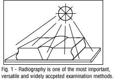
Radiography (X-ray) is one of the most important, versatile and widely accepted of all the nondestructive examination methods - Fig. 1. X-ray is used to determine internal soundness of the welds. The term "X-ray quality," widely used to indicate high quality in welds, arises from this inspection method.
Radiography is based on the ability of X-rays and gamma rays to pass through metal and other materials opaque to ordinary light, and produce photographic records of the transmitted radiant energy. All materials will absorb known amounts of this radiant energy and, therefore, X-rays and gamma rays can be used to show discontinuities and inclusions within the opaque material. The permanent film record of the internal conditions will show the basic information by which weld soundness and be determined.
X-rays are produced by high-voltage generators. As the high voltage applied to an X-ray tube is increased, the wavelength of the emitted X-ray becomes shorter , providing more penetrating power. Gamma rays are produced by the atomic disintegration of radioisotopes. The radioactive isotopes most widely used in industrial radiography are Cobalt 60 and Iridium 192. Gamma rays emitted from these isotopes are similar to X-rays, except their wavelengths are usually shorter. This allows them to penetrate to greater depths than X-rays of the same power, however, exposure times are considerably longer due to the longer intensity.
When X-rays or gamma rays are directed at a section of weldment , not all of the radiation passes are through the metal. Different materials, depending on their density, thickness and atomic number, will absorb different wavelengths of radiant energy.
The degree to which the different materials absorb these rays determines the intensity of the rays penetrating through the material. When variations of these rays are recorded, a means of seeing inside the material is available. The image on a developed photo-sensitized film is known as a radiograph. Thicker areas of the specimen or higher density material (tungsten inclusion), will absorb more radiation and their corresponding areas on the radiograph will be lighter - Fig 2.
Whether in the shop or in the field, the reliability and interpretive value of radiographic images are a function of their sharpness and contrast. The ability of an observer to detect a flaw depends on the sharpness of its image and its contrast with the background. To be sure that a radiographic exposure produces acceptable results, a gauge known as an Image Quality Indicator (IQI) is placed on the part so that its image will be produced on the radiograph.
IQI's used to determine radiographic quality are also called penetrameters. A standard hole-type penetrameter is a rectangular piece of metal with three drilled holes of set diameters. The thickness of the piece of metal is a percentage of the thickness of the specimen being radiographed. The diameter of each hole is different and is a given multiple of the penetrameter thickness. Wire-type penetrameters are also widely used, especially outside the United States. They consist of several pieces of wire, each of a different diameter. Sensitivity is determined by the smallest diameter of wire that can be clearly seen on the radiograph.
A penetrameter is not an indicator or gauge to measure the size of a discontinuity or the minimum detectable flaw size. It is an indicator of the quality of the radiographic technique.
Radiographic images are not always easy to interpret. Film handling marks and streaks, fog and spots caused by developing errors may make it difficult to identify defects. Such film artifacts may mask weld discontinuities.
Surface defects will show up on the film and must be recognized. Because the angle of exposure will also influence the radiograph, it is difficult or impossible to analyze fillet welds by this method. Because a radiograph compresses all the defects that occur throughout the thickness of the weld into one plane, it tends to give an exaggerated impression of scattered type defects such as porosity or inclusions.
An X-ray image of the interior of the weld may be viewed on a fluorescent screen, as well as on developed film. This makes it possible to inspect parts faster and at a lower cost, but the image definition is poorer. Computerization has made it possible to overcome many of the shortcomings of radiographic imaging by linking the fluorescent screen with a video camera. Instead of waiting for film to be developed, the images can be viewed in real time. This can improve quality and reduce costs on production applications such as pipe welding, where a problem can be identified and corrected quickly.
By digitizing the image and loading it into a computer, the image can be enhanced and analyzed to a degree never before possible. Multiple images can be superimposed. Pixel values can be adjusted to change shading and contrast, bringing out small flaws and discontinuities that would not show up on film. Colors can be assigned to the various shades of gray to further enhance the image and make flaws stand out better. The process of digitizing an image taken from the fluorescent screen - having that image computer enhanced and transferred to a viewing monitor - takes only a few seconds. However, because there is a time delay, we can no longer consider this "real time." It is called "radioscopy imagery."
Existing films can be digitized to achieve the same results and improve the analysis process. Another advantage is the ability to archive images on laser optical disks, which take up far less space than vaults of old films and are much easier to recall when needed.
Industrial radiography, then, is an inspection method using X-rays and gamma rays as a penetrating medium, and densitized film as a recording medium, to obtain a photographic record of internal quality. Generally, defects in welds consist either of a void in the weld metal itself or an inclusion that differs in density from the surrounding weld metal.
Radiographic equipment produces radiation that can be harmful to body tissue in excessive amounts, so all safety precautions should be followed closely. All instructions should be followed carefully to achieve satisfactory results. Only personnel who are trained in radiation safety and qualified as industrial radiographers should be permitted to do radiographic testing.
Magnetic Particle Inspection (MT)
Magnetic particle inspection is a method of locating and defining discontinuities in magnetic materials. It is excellent for detecting surface defects in welds, including discontinuities that are too small to be seen with the naked eye, and those that are slightly subsurface.
This method may be used to inspect plate edges prior to welding, in process inspection of each weld pass or layer, postweld evaluation and to inspect
repairs - Fig. 3.
It is a good method for detecting surface cracks of all sizes in both the weld and adjacent base metal, subsurface cracks, incomplete fusion, undercut and inadequate penetration in the weld, as well as defects on the repaired edges of the base metal. Although magnetic particle testing should not be a substitute for radiography or ultrasonics for subsurface evaluations, it may present an advantage over their methods in detecting tight cracks and surface discontinuities.
With this method, probes are usually placed on each side of the area to be inspected, and a high amperage is passed through the workplace between them. A magnetic flux is produced at right angles to the flow of current - Fig. 3. When these lines of force encounter a discontinuity, such as a longitudinal crack, they are diverted and leak through the surface, creating magnetic poles or points of attraction. A magnetic powder dusted onto the surface will cling to the leakage area more tenaciously than elsewhere, forming an indication of the discontinuity.
For this indication to develop, the discontinuity must be angled against the magnetic lines of force. Thus, when current is passed longitudinally through a workpiece, only longitudinal flaws will show. Putting the workpiece inside a solenoid coil will create longitudinal lines of force (Fig. 3) that cause transverse and angular cracks to become visible when the magnetic powder is applied.
Although much simpler to use than radiographic inspection, the magnetic particle method is limited to use with ferromagnetic materials and cannot be used with austenitic steels. A joint between a base metal and a weld of different magnetic characteristics will create magnetic discontinuities that may falsely be interpreted as unsound. On the other hand, a true defect can be obscured by the powder clinging over the harmless magnetic discontinuity. Sensitivity decreases with the size of the defect and is also less with round cracks such as gas pockets. It is best with elongated forms, such as cracks, and is limited to surface flaws and some subsurface flaws, mostly on thinner materials.
Because the field must be distorted sufficiently to create the external leakage required to identify flaws, the fine, elongated discontinuities, such as hairline cracks, seams or inclusions that are parallel to the magnetic field, will not show up. They can be developed by changing the direction of the field, and it is advisable to apply the field from two directions, preferably at right angles to each other.
Magnetic powders maybe applied dry or wet. The dry powder method is popular for inspecting heavy weldments, while the wet method is often used in inspecting aircraft components. Dry powder is dusted uniformly over the work with a spray gun, dusting bag or atomizer. The finely divided magnetic particles are coated to increase their mobility and are available in gray, black and red colors to improve visibility. In the wet method, very fine red or black particles are suspended in water or light petroleum distillate. This can be flowed or sprayed on, or the part may be dipped into the liquid. The wet method is more sensitive than the dry method, because it allows the use of finer particles that can detect exceedingly fine defects. Fluorescent powders may be used for further sensitivity and are especially useful for locating discontinuities in corners, keyways, splines and deep holes.
Liquid Penetrant Inspection (PT)
Surface cracks and pinholes that are not visible to the naked eye can be located by the liquid penetrant inspection. It is widely used to locate leaks in welds and can be applied with austentic steels and nonferrous materials where magnetic particle inspection would be useless.
Liquid penetrant inspection is often referred to as an extension of the visual inspection method. Many standards, such as the AWS D.1. Code, say that "welds subject to liquid penetrant testing�shall be evaluated on the basis of the requirements for visual inspection."
Two types of penetrating liquids are used - fluorescent and visible dye. With fluorescent penetrant inspection, a highly fluorescent liquid with good penetrating qualities is applied to the surface of the part to be examined. Capillary action draws the liquid into the surface openings, and the excess is then removed. A "developer" is used to draw the penetrant to the surface, and the resulting indication is viewed by ultraviolet (black) light. The high contrast between the fluorescent material and the object makes it possible to detect minute traces of penetrant that indicate surface defects.
Dye penetrant inspection is similar, except that vividly colored dyes visible under ordinary light are used - Fig. 4. Normally, a white developer is used with the dye penetrants that creates a sharply contrasting background to the vivid dye color. This allows greater portability by eliminating the need for ultraviolet light.
The part to be inspected must be clean and dry, because any foreign matter could close the cracks or pinholes and exclude the penetrant. Penetrants can be applied by dipping, spraying or brushing, but sufficient time must be allowed for the liquid to be fully absorbed into the discontinuities. This may take an hour or more in very exacting work.
Liquid penetrant inspection is widely used for leak detection. A common procedure is to apply fluorescent material to one side of a joint, wait an adequate time for capillary action to take place, and then view the other side with ultraviolet light. In thin-walled vessels, this technique will identify leaks that ordinarily would not be located by the usual air test with pressures of 5-20 lb/in.2 When wall thickness exceeds � in., however, sensitivity of the leak test decreases.
Ultrasonic Inspection (UT)
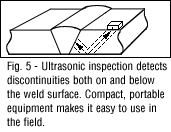
Ultrasonic inspection is a method of detecting discontinuities by directing a high-frequency sound beam through the base plate and weld on a predictable path. When the sound beam's plate path strikes an interruption in the material continuity, some of the sound is reflected back. The sound is collected by the instrument, amplified and displayed as a vertical trance on a video screen - Fig. 5.
Both surface and subsurface detects in metals can be detected, located and measured by ultrasonic inspection, including flaws too small to be detected by other methods.
The ultrasonic unit contains a crystal of quartz or other piezoelectric material encapsulated in a transducer or probe. When a voltage is applied, the crystal vibrates rapidly. As an ultrasonic transducer is held against the metal to be inspected, it imparts mechanical vibrations of the same frequency as the crystal through a couplet material into the base metal and weld. These vibrational waves are propagated through the material until they reach a discontinuity or change in density. At these points, some of the vibrational energy is reflected back. As the current that causes the vibration is shut off and on at 60-1000 times per second, the quartz crystal intermittently acts as a receiver to pick up the reflected vibrations. These cause pressure on the crystal and generate an electrical current. Fed to a video screen, this current produces vertical deflections on the horizontal base line. The resulting pattern on the face of the tube represents the reflected signal and the discontinuity. Compact portable ultrasonic equipment is available for field inspection and is commonly used on bridge and structural work.
Ultrasonic testing is less suitable than other NDE methods for determining porosity in welds, because round gas pores respond to ultrasonic tests as a series of single point reflectors. This results in low-amplitude responses that are easily confused with "base-line noise" inherent with testing parameters. However, it is the preferred test method for detecting plainer-type discontinuities and lamination.
Portable ultrasonic equipment is available with digital operation and microprocessor controls. These instruments may have built-in memory and can provide hard-copy printouts or video monitoring and recording. They can be interfaced with computers, which allows further analysis, documentation and archiving, much as with radiographic data. Ultrasonic examination requires expert interpretation from highly skilled and extensively trained personnel.
Choices Control Quality
A good NDE inspection program must recognize the inherent limitations of each process. For example, both radiography and ultrasound have distinct orientation factors that may guide the choice of which process to use for a particular job. Their strengths and weaknesses tend to compliment each other. While radiography is unable to reliably detect lamination-like defects, ultrasound is much better at it. On the other hand, ultrasound is poorly suited to detecting scattered porosity, while radiography is very good.
Whatever inspection techniques are used, paying attention to the "Five P's" of weld quality will help reduce subsequent inspection to a routine checking activity. Then, the proper use of NDE methods will serve as a check to keep variables in line and weld quality within standards.
The Five P's are:
1. Process Selection - the process must be right for the job.
2. Preparation - the joint configuration must be right and compatible with the welding process.
3. Procedures - the procedures must be spelled out in detail and followed religiously during welding.
4. Pretesting - full-scale mockups or simulated specimens should be used to prove that the process and procedures give the desired standard of quality.
5. Personnel - qualified people must be assigned to the job.